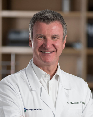
D. Geoffrey Vince Laboratory
-
D. Geoffrey Vince Laboratory
- Principal Investigator
- Research
- Our Team
- Publications
- Patents
- Research News
Research
Dr. Vince no longer maintains an active research laboratory. This page reflects his leadership role within Cleveland Clinic.
D. Geoffrey Vince, PhD, is a nationally recognized leader in healthcare innovation, serving as Executive Director at Cleveland Clinic Innovations and Chair of Biomedical Engineering at Cleveland Clinic Research. He holds The Virginia Lois Kennedy Chair in Biomedical Engineering and Applied Therapeutics.
Dr. Vince focuses on aligning commercialization efforts with research priorities, fostering growth in data and computing sciences and accelerating healthcare technology and therapy innovations. His influence extends to Cleveland Clinic’s Board of Governors, and he has been named among Becker’s "Top 35 Chief Innovation Officers to Know" for three consecutive years.
Biography
Dr. Vince earned his PhD in biomedical engineering from the University of Liverpool and joined Cleveland Clinic in 1992 as a Postdoctoral Research Fellow, becoming Associate Staff by 2003. During this time, he co-developed the novel "Virtual Histology™" technology for imaging coronary arteries using intravascular ultrasound, a Cleveland Clinic innovation that was patented and licensed to Volcano Therapeutics (now part of Philips).
To bring this technology to market, Dr. Vince took on leadership roles at Volcano Therapeutics between 2005 and 2011, first as Director of Research and later as Vice President of Clinical and Advanced R&D. Under his guidance, Virtual Histology™ became a globally recognized product, with approximately 7,000 units deployed worldwide. This achievement is just one example of his prolific work as an inventor, with more than 60 patents worldwide.
In 2011, Dr. Vince returned to Cleveland Clinic to chair the Department of Biomedical Engineering, continuing to advance innovation and commercialization efforts. In 2022, he was appointed Executive Director of Cleveland Clinic Innovations. He focuses on aligning commercialization efforts with research priorities, fostering growth in data and computing sciences, and accelerating healthcare technology and therapy innovations.
Dr. Vince recently served as Principal Investigator for Cleveland Clinic in an NIH Centers for Accelerated Innovations grant, aimed at translating biomedical advances into commercially viable products that enhance patient care and public health.
Dr. Vince's work has advanced biomedical engineering while driving economic growth in Northeast Ohio. His leadership in healthcare innovation has earned him recognition on Becker's Healthcare's "Top 35 Hospital and Health System Chief Innovation Officers to Know" list for three consecutive years.
Education & Professional Highlights
Fellowship - Cleveland Clinic
Biomedical Engineering
Cleveland, OH USA
1995
Medical Education - University of Liverpool Faculty of Medicine
Clinical Engineering
Liverpool, UK
1989
Undergraduate - Leicester DeMontford University
Medical Sciences & Chemistry
Leicester, UK
1985
Research
Dr. Vince previously led a research laboratory but now serves exclusively in a leadership/administrative role and does not conduct active lab research.
Our Team
Selected Publications
View publications for D. Geoffrey Vince, PhD
(Disclaimer: This search is powered by PubMed, a service of the U.S. National Library of Medicine. PubMed is a third-party website with no affiliation with Cleveland Clinic.)
Patents
| US Patent | Patent Title | Issue Date | First-Named Inventor |
|---|---|---|---|
| 8,630,492 | System and Method for Identifying a Vascular Border | 01/14/2014 | D. Geoffrey Vince, PhD |
| 8,622,910 | System and Method of Acquiring Blood-Vessel Data | 01/04/2014 | D. Geoffrey Vince, PhD |
| 6,200,268 | Vascular Plaque Characterization | 03/13/2001 | D. Geoffrey Vince, PhD |
Research News

Dr. Vince, an inventor and scientist, leads Cleveland Clinic Innovations and chairs the Department of Biomedical Engineering.

Dr. Vince will lead the commercialization arm of Cleveland Clinic, which turns medical breakthrough inventions into patient-benefiting medical products and companies.

With a new award from the Department of Defense, Dr. Vince will use non-invasive ultrasound and a novel artificial intelligence algorithm that predicts carotid artery plaque composition to detect patients at high risk of future stroke.
